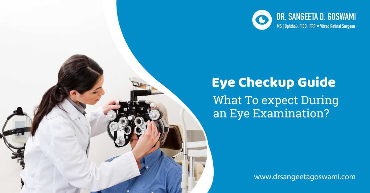

A standard eye checkup takes less than an hour but covers everything from visual clarity to eye disease screening.
To help you understand things better, this guide explains what to expect during an eye examination.
Our eyes are under constant strain from prolonged screen time, environmental factors, ageing, and underlying health conditions.
These can include diabetes or high blood pressure, which can affect your vision, so regular eye examinations are essential for protecting your vision at any age.
You can consult the best eye doctor in Siliguri to schedule your comprehensive eye test and eliminate any early signs of defects in your eyes.
A comprehensive eye examination is a multi-step process that evaluates both your vision and overall ocular health.
These tests typically begin with a medical history review, followed by a visual acuity test, refraction, a slit lamp exam, and a check for glaucoma, lasting between 30 and 60 minutes.
What is an Eye Checkup?
A routine medical appointment where an eye specialist evaluates both your visual clarity and the physical health of your eyes is called an eye checkup.
To detect vision changes and hidden eye diseases long before symptoms appear, it combines simple sight tests with advanced structural exams.
Here is what happens during these visits.
- Vision Examination: The first thing done in a comprehensive eye test is your vision examination. Here you read letters from a chart to see if you need glasses or a prescription update.
- Eye Muscle Test:The eye muscle test is done by the doctor tracking your eye movements to ensure your eyes work together correctly.
- Fluid Pressure Test: The fluid pressure test is done to screen for glaucoma, with aquick air puff or probe, which measures internal eye pressure.
- Microscopic Inspection or Examination: The microscopic inspection or examination is done with high-powered lights that magnify the front and back structures of your eye to check for cell damage.
When Should You Get an Eye Exam Done?
These scheduled tests are important for prevention and early diagnosis because many eye disorders, including glaucoma, cataracts, and retinal diseases, do not show early symptoms.
1. Catching Silent Early Eye Diseases
These silent eye diseases can be.
- Glaucoma:
Characterized by irreversible damage to the optic nerve, which is the vital cable transmitting visual data from your eye to your brain. Glaucoma is a group of progressive eye diseases.
It typically develops painlessly and gradually destroys the optic nerve due to high internal fluid pressure before any clear warning signs appear, and is often called the “silent thief of sight”.
- Macular Degeneration:
Macular degeneration is a common eye disease that damages the macula, which is the central part of the retina responsible for sharp, detailed vision. It also gradually destroys sharp, central vision required for reading and driving.
It primarily affects older adults, causing blurred central vision or blind spots, while leaving peripheral or side vision completely intact.
- Cataracts:
A cataract is a clouding of the eye’s natural lens, which causes blurred vision, faded colors, and sensitivity to glare.
It is primarily caused by aging as proteins in the lens break down and clump together.
The condition is highly treatable and is permanently corrected through a quick, outpatient surgical procedure.
2. Identifying Generalized Health Issues
Eye tests can help in identifying generalized health issues as well.
Because the eyes are the only place in your body where a doctor can see blood vessels, connective tissues, and nerves directly without surgery.
These exams can help determine early signs of the following.
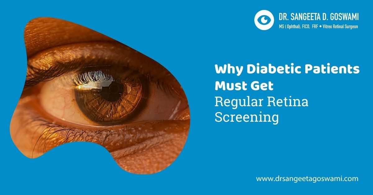
- Diabetes:
By damaging the microvascular system, which is the network of tiny blood vessels, throughout the entire body, diabetes causes generalized or systemic eye health issues.
It causes tiny retinal blood vessels to leak fluid or bleed, called diabetic retinopathy.
- High Blood Pressure:
High blood pressure, or systemic hypertension, impacts your eye health by forcing high-pressure blood flow through the delicate microvascular network of the eye.
Over time, this mechanical stress disrupts blood flow, damages the vessel walls, and alters the structures responsible for crisp vision. Also forces blood vessels to narrow, twist, or burst.
- High Cholesterol:
Hypercholesterolemia or high cholesterol impacts the eyes through two mechanisms: atherosclerotic blockages within the retinal blood supply and the structural deposition of lipids in ocular tissues.
Because excess lipids circulate throughout the entire bloodstream, they leave distinct physical and vascular markers inside and around the eye structures.
This creates a distinctive yellowish or blue ring around the cornea or visible plaques in retinal arteries.
3. Preventing Chronic Fatigue and Eye Strain
Eye tests also help in preventing chronic fatigue and eye strain because outdated prescriptions or uncorrected minor vision changes force your brain and eye muscles to work harder. And scheduling regular checkups can eliminate the following.
- Frequent headaches
- Dry, burning, or watery eyes
- Blurred vision while working on computers or driving at night.

Eye Checkup Guide: What To Expect During an Eye Examination?
Evaluating both your clarity of vision and your structural eye health, a comprehensive eye examination is a multi-step checkup.
In these exams, an eye care specialist uses specialized equipment to screen for refractive errors and early signs of silent eye diseases, and the entire appointment generally lasts about an hour.
Here are the steps that an expert includes during an eye examination.
1. Step 1- Learning the Patient’s History and Pre-Screening
The first step to conducting an eye exam starts with the doctor learning about your history and starting the pre-screening of your eyes.
- Medical Review:Then comes the medical review by the doctors, which is where they discuss your general health, active medications, and family history of chronic issues like diabetes or high blood pressure.
- Discussion of Your Symptoms: Based on your medical review, there will be a discussion of the symptoms that you have.Here you will have to mention any recent vision changes, dry eye irritation, or work environment strains.
2. Step 2- Visual Acuity and Alignment Tests
The second step to conducting your eye exam is to check for visual acuity and alignment tests, where your doctor measures the absolute sharpness of your vision and checks how well your eye muscles work together.
These tests include the following.
- Snellen Chart: TheSnellen chart is used in eye tests by making you read rows of progressively smaller letters from a fixed distance to measure your visual sharpness.
- Muscle Tracking: The muscle tracking is a test in which the specialist evaluates your ocular motility by moving an object or penlight to ensure your eyes coordinate properly.
- Cover Test: In a cover test, your eyes are covered momentarily to observe how the uncovered eye moves to adjust focus, checking for eye alignment problems.
3. Step 3- Conducting Refraction Testing
The third step in conducting your eye examination is refraction testing, where an eye care professional determines the precise lens power you need to achieve 20\20 vision.
This step identifies whether you have a refractive error, meaning the shape of your eye bends light incorrectly, and calculates your prescription for glasses or contact lenses.
- Phoropter Fine-Tuning:During the phoropter fine-tuning, you will have to look through a phoropter device, done for patients who experience blurry vision.
- Lens Comparison:Lens comparison is a small test, where the doctor switches back and forth between different lens powers, asking you which option provides the sharpest view.
- Eyewear Prescription:The final data determines your correct prescription strength for glasses or contact lenses. On the basis of the previous tests, the doctor then prescribes the necessary lens needed for the eyes.
4. Step 4- Glaucoma Screening or Intraocular Pressure
The fourth step of your eye exam is conducting the Glaucoma Screening or Intraocular Pressure (IOP) test, where the doctor measures the fluid pressure inside your eyes.
This is a critical screening tool for glaucoma, a group of eye diseases that damage the optic nerve and cause permanent vision loss, often with zero early warning signs or pain.
- Tonometry:Tonometry is a medical screening that calculates the fluid pressure inside your eyeball.
- Air Puff or Contact: A machine will puff a quick burst of air or gently tap the surface using a specialized sensor.
- Glaucoma Warning:Elevated pressure is a major, often symptomless warning indicator for irreversible optic nerve damage.
5. Step 5- Conducting the Front-of-Eye Evaluation or Slit Lamp
In this step, the doctor uses a specialized microscope called a slit lamp to examine the physical structures at the front of your eye.
Allowing the specialist to view your anatomy in clear, three-dimensional cross-sections, this high-magnification tool projects a thin, intense slice of light, a slit, into the eye.
- Biomicroscope Examination: In biomicroscopic examination, you place your chin on a rest while a high-magnification microscope illuminates the eye.
- Anterior Anatomy: In this, the doctor checks the health of your eyelids, cornea, conjunctiva, iris, and crystalline lens.
- Cataract Screening:This specific test detects the presence of physical lens clouding or ocular surface damage.
6. Step 6- Back-of-Eye Assessment: A Retinal Exam
The sixth step of conducting an eye exam is conducting the back-of-eye assessment, which is a retinal exam, where your doctor examines the interior chamber of your eye.
Offering crucial clues about both your eyesight and your systemic health, this test allows direct visualization of live blood vessels, the retina, and the optic nerve without surgery.
- Dilating Drops:Here, special drops are frequently applied to widen your pupils completely, taking about 30 minutes to work.
- Fundoscopy View:In fundoscopy view, the doctor uses an ophthalmoscope to inspect the back of your eye cavity.
- Deep Tissue Scan:During a deep tissue scan, the doctor looks closely at the retina, macula, and the optic nerve connection.
- Systemic Indicators:This internal check can reveal early microvascular signs of diabetes, high cholesterol, and brain anomalies.
Some Important Preparation Tips
1. Bring Eyewear to Your Test:
To check the old prescription accuracy, bring your current glasses, contact lenses, or over-the-counter reading specs. This will help make sure that you are applying and using the right and prescribed medicines.
2. Plan Your Ride Home:
Pupil dilation causes blurry near vision and high light sensitivity for roughly 4 to 6 hours. So it is best to plan your ride home safely by either asking your loved ones to drop you home or taking a rental car ride home from the clinic.
3. Packing Sunglasses is Essential:
Bring dark sunglasses to protect your dilated pupils from solar glare directly after your appointment, which is essential after your treatment, as your eyes should not come in contact with the sun.
Conclusion
A comprehensive eye examination is a painless, multstep health checkup that evaluates both your visual clarity and the physical health of your eyes, typically lasting up to 30 to 60 minutes.
There are many steps in conducting a successful eye examination, some of which are learning the patient’s history, visual acuity and alignment tests, pre- screenings, glaucoma screening, intraocular pressure, and many more.
Consult the best eye doctor in Siliguri to schedule your comprehensive eye test and eliminate any early signs of defects in your eyes.
You can secure an appointment with Dr. Sangeeta D. Goswami to conduct the best eye examination and detect any early signs of defects in your eyes.